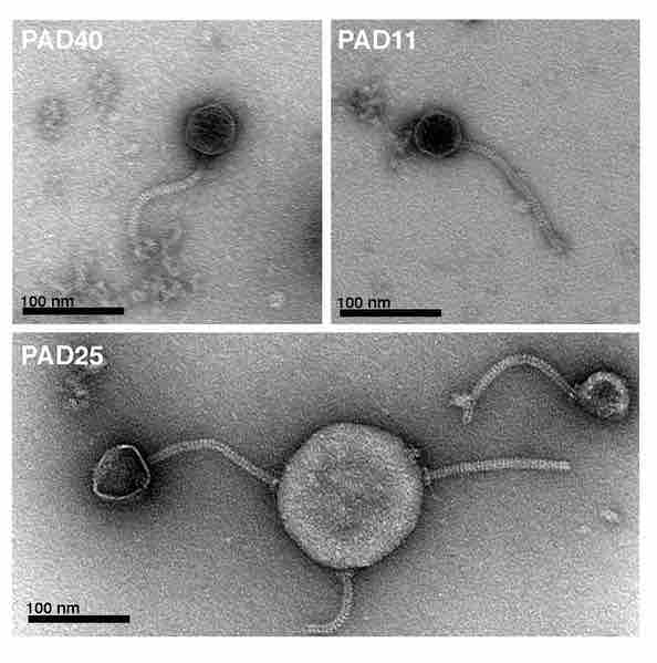
Siphovirus phages
Electron micrographs of bacteriophages from P. acnes. Phages were negatively stained with 0.75% uranyl formate and subjected to transmission electron microscopy. The phages have a head of approximately 55 nm in diameter, loaded with genetic material. Their tails have a size of 150 × 10 nm and are flexible and non-contractile. In the lower micrograph, PAD25 is adhering to bacterial cell debris, and two phages have lost their heads. At the attachment site between the phage and the cell debris, a base plate with attached spikes can be observed. All phages were classified as Siphoviruses based on their morphology.
Source
Boundless vets and curates high-quality, openly licensed content from around the Internet. This particular resource used the following sources: